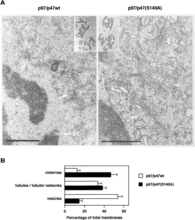
Figure 8.

The microinjection of p47(S140A) preserves Golgi stacks in mitotic cells. (A) Representative EM images of Golgi at late prometaphase in the cells injected with either p97/p47wt or p97/p47(S140A) as described in Fig. 7 A. Insets show the whole cell images. White arrows show Golgi. Bars, 1 μm. (B) In the injected cells at prometaphase, Golgi membranes were classified and counted as described previously (Shorter and Warren, 1999; Seemann et al., 2000). Means ± SE (n = 9).
