
Figure 2.
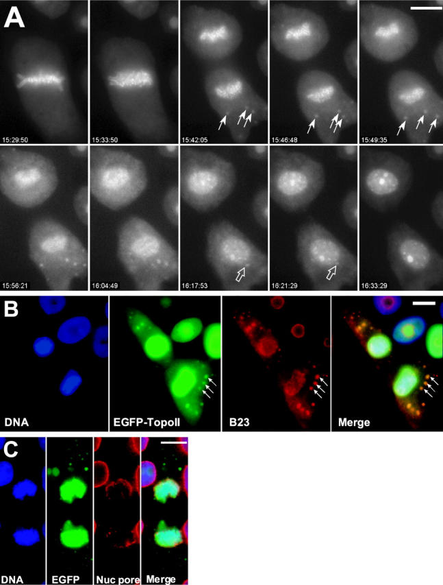
EGFP–topoIIα localization is dynamic at anaphase and telophase. (A) Selected frames from a time-lapse video of a GT2-LPk cell in late mitosis. In anaphase, some of the cytoplasmic EGFP–topoIIα concentrates into distinct foci (closed arrows) that are motile. At telophase, the foci appear to fuse with the reforming nucleus (open arrows), whereas nucleoli become more apparent. (Image frames in this sequence were individually adjusted for contrast to optimize visualization of the cyto- plasmic foci; Video 1, available at http://www.jcb.org/cgi/content/full/jcb.200202053/DC1) (B) EGFP–topoIIα foci (arrows) in telophase cells colocalize with NDFs identified by labeling with antibody to nucleolar B23 protein, a gift from Dr. Mirek Dundr (National Cancer Institute, Bethesda, MD). (The EGFP– topoIIα image is oversaturated for the nuclear fluorescence to clearly visualize the cytoplasmic foci.) (C) EGFP–topoIIα foci do not colocalize with reforming nuclear envelope identified by RL1 antibody to nuclear pore proteins, a gift from Dr. Bryce Pascal (University of Virginia, Charlottesville, VA). Bars, 10 μm.