
Figure 5.
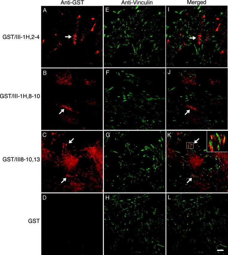
Localization of III1H constructs on cell surfaces. Collagen-adherent Fn−/− cells were incubated with 250 nM of GST–III1H,2–4 (A, E, and I), GST–III1H,8–10 (B, F, and J), GST–III8–10,13 (C, G, and K), or GST (D, H, and L). Cells were processed for immunofluorescence as indicated in the Materials and methods. Fusion proteins were visualized using a polyclonal anti-GST antibody followed by a Texas red–labeled goat anti–rabbit antibody (A–D). Vinculin was visualized using an antivinculin mAb followed by an FITC-labeled goat anti–mouse antibody (E–H). In the merged images shown in I–L, areas of colocalization appear yellow. Bar, 10 μM.