Figure 3.
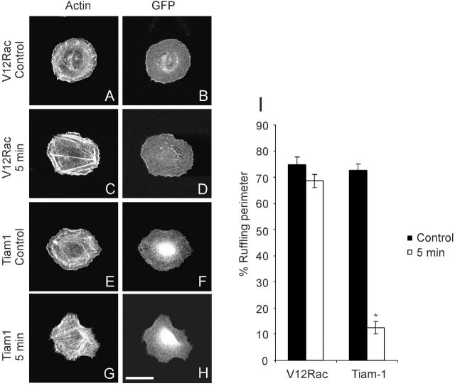
Effects of stretch on V12Rac- and Tiam1-transfected VSM cells. (A–D) VSM cells were transfected with 2 μg of pEGFP-C1-V12Rac. (E–H) VSM cells were cotransfected with 1.6 μg of pcDNAINeo-Tiam1 and 0.4 μg of pEGFP. At 24 h after transfection, cells were plated on collagen I–coated silicone membrane for 3 h and stretched equibiaxially by 15% for 5min. Fluorescence images of actin filaments (A, C, E, and G) and GFP (B, D, F, and H) in the same cells were shown. Results are representative of three experiments. Bar, 20 μm. (I) The portion of the cell perimeter occupied by lamellipodia is expressed as percent of the total perimeter. Values are means ± SEM, for 20 cells per data point. * indicates P < 0.01.

