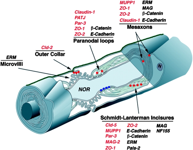
Figure 11.
Location of autotypic junction proteins in myelinating Schwann cells. The organization of peripheral myelinated nerve is shown schematically. The location of autotypic junctions in the Schmidt Lanterman incisures, paranodal loops, mesaxons and the outer aspect of the nodal gap are marked with red dots. The heterotypic septate-like junction formed between the axon and the paranodal loops of the myelinating Schwann cell is labeled with blue dots. The basal lamina covering the Schwann cell-axon unit is shown in green. The localization of different proteins discussed in this study is listed. Tight junction proteins are labeled in red. Note that autotypic tight junctions present in different aspects of noncompact myelin contain distinct junctional complexes (modified from Spiegel and Peles [2002] and used with permission of Taylor and Francis, www.tandf.co.uk).

