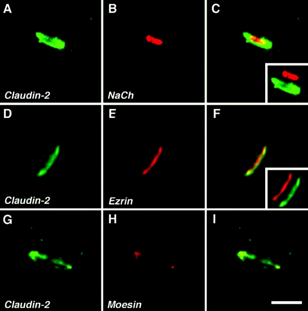
Figure 7.
Localization of claudin-2 to the nodal region in peripheral nerves. Images of teased fibers from adult mouse sciatic nerve, double labeled for claudin-2 (green) and voltage-gated Na+ channels (A–C, NaCh), ezrin (D–F), or moesin (G–I), all in red. The left panel of each row shows the merged images. The insets in C and F show merged images in which the red and the green channels were shifted apart. Note that nodal membranes were labeled for NaCh, and were surrounded by a ring of claudin-2 staining. Claudin-2 colocalized with ezrin and moesin, which labeled Schwann cell microvilli. Bar, 5 μm.

