
Figure 5.
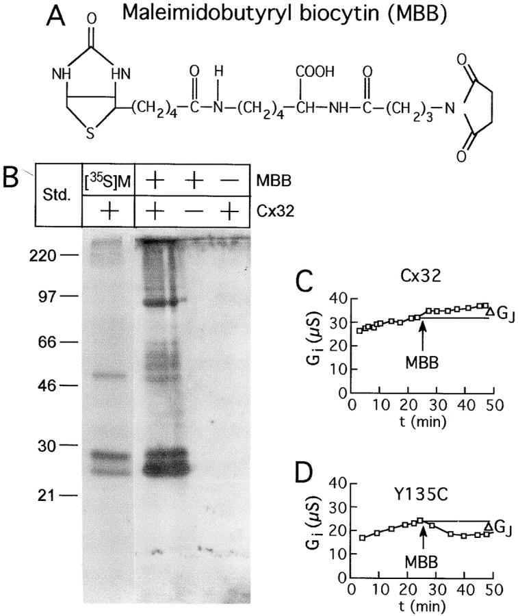
Structure of the thiol reagent maleimidobutyryl biocytin (MBB), and detection of its reactivity with Cx32 in the perfusion chamber. (A) Irreversible reaction occurs between the SH group of a cysteine and the maleimide ring, with the biotin moiety providing easy detection. (B) Biochemical determination that MBB reacts with Cx32 in the dual-oocyte perfusion system. Radiolabeled Cx32, immunoprecipitated from intact oocytes, shows a full-length and partially degraded form just below the 30-kD marker, and an aggregated dimer at ∼50 kD (left lane). Cx32 detected by avidin blot after intracellular perfusion of a paired oocyte with 1 mM MBB and immunoprecipitation of Cx32 (second lane from left) reveals the same bands, although the increased sensitivity of avidin detection revealed additional aggregated forms, both dimeric (50–60 kD) and trimeric (∼90 kD). No protein is detected for noninjected oocytes exposed to MBB or for perfused Cx32-injected oocytes not exposed to MBB (left two lanes). (C and D) Changes in junctional conductance (Gj) during typical SCAM experiments. In most cases, conductance increased steadily before and after addition of the reagent as shown for wtCx32 (C). In cases where a substituted cysteine was accessible, a decrease in conductance occurred after the addition of MBB, as shown for Cx32Y135C (D). The effect of MBB on each oocyte pair was determined by comparison of the conductance immediately before addition of the reagent, to that 20 min after its application (ΔGj).