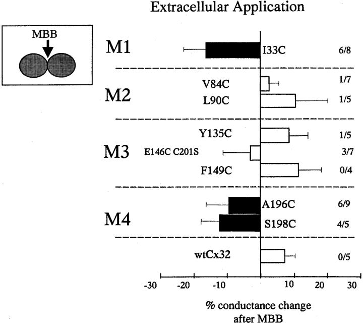
Figure 7.
Changes in conductance after extracellular MBB application to intact oocyte pairs where access to the pore was not possible. As in the perfusion experiments in Fig. 6, wtCx32 and cysteine substitution mutants in M2 and M3 continued to show small increases in conductance after addition of the reagent. In contrast, significant decreases in conductance compared to wt (P ≤ 0.05; t test) were observed after application of MBB to a number of cysteine substitution mutants in M1 and M4 (filled bars). In all of these cases, block was also observed in >50% of perfusions. Reactivity suggests that the designated cysteines lie within an MBB-accessible environment, continuous with the extracellular space but not in the pore. Data are presented in the same format as Fig. 6.

