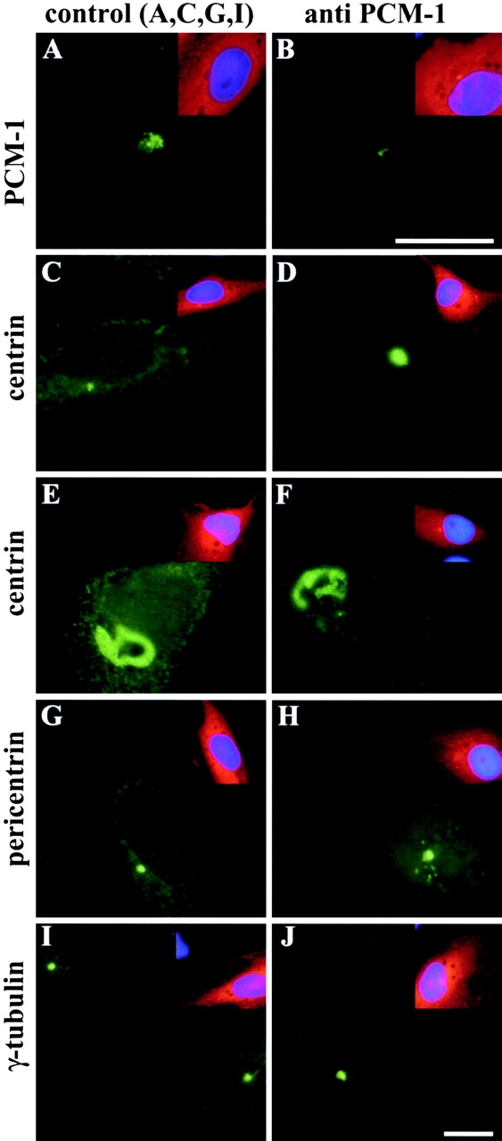
Figure 2.

Microinjection of antibodies against PCM-1 causes aggregation of centrin and pericentrin. Xenopus A6 cells were microinjected with affinity-purified antibodies against PCM-1 (B, D–F, H, and J), or with control antibodies (A, C, G, and I). Cells were stained for immunofluorescence of (A and B) PCM-1, (C–F) centrin, (G and H) pericentrin, or (I and J) γ-tubulin. Insets show the same cells stained with Texas red–labeled anti–rabbit antibody to identify microinjected cells, and stained with DAPI to detect DNA (blue). Bars: (B and J) 10 μm; same magnifications in A and B and C–J, respectively.
