Figure 5.
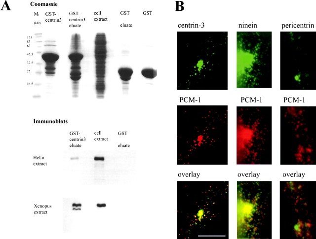
PCM-1 binds to centrin, and partly colocalizes with centriolar satellites of centrin, ninein, and pericentrin. (A) Binding assay using glutathione beads on cell extracts preincubated with GST-tagged centrin-3 or with GST alone. Lanes on a Coomassie-stained gel show relative molecular mass markers (Mr), purified GST-centrin-3, the eluate from glutathione beads incubated with Xenopus egg extract and GST-centrin-3, egg extract alone, the eluate from glutathione beads incubated with egg extract and GST, and purified GST alone. Positions of molecular weight markers are indicated on the left. Shown below are immunoblots of corresponding lanes, probed with antibodies against PCM-1. (B) Immunofluorescence staining of centriolar satellites. Left column, confocal section of a PtK2 cell stained for centrin-3 (green) and PCM-1 (red). Middle and right columns, conventional immunofluorescence of mouse myoblast cells stained for (middle) ninein and PCM-1, or (right) pericentrin and PCM-1. Bar (B), 10 μm.

