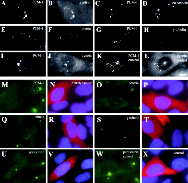
Figure 6.
Microtubules and dynactin are essential for the centrosomal accumulation of PCM-1, centrin, pericentrin, and ninein, but not γ-tubulin. (A–J) Image pairs of CHO cells after nocodazole treatment are shown, stained for (A and B) PCM-1 and centrin, (C and D) PCM-1 and pericentrin, (E and F) PCM-1 and ninein, (G and H) PCM-1 and γ-tubulin, and (I and J) PCM-1 and dynein intermediate chain. K and L show an untreated cell stained for PCM-1 and dynein intermediate chain. Arrowheads indicate pericentriolar dynein spots colocalizing with PCM-1 granules. (M–V) Image pairs of CHO cells microinjected with p50/dynamitin are shown. (M, O, Q, S, and U) Cells were stained for PCM-1, centrin-3, ninein, γ-tubulin, and pericentrin, respectively. (N, P, R, T, and V) Corresponding images showing dynamitin-injected cells (red) and DNA staining (blue). Dynamitin-dependent inhibition of centrosomal localization varied for different proteins; PCM-1 dispersed in 77% of injected cells (n = 106, controls 2%, n = 182), centrin was affected in 60% (n = 50, controls 2%, n = 191), ninein in 45% (n = 110, controls 3%, n = 169), pericentrin in 33% (n = 470, controls 5%, n = 73),and γ-tubulin in 3% (n = 76, controls 1%, n = 135). (W and X) Image pair of a control cell injected with labeled goat anti–rabbit antibody and stained for pericentrin. Bar (L), 10 μm.

