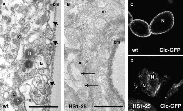
Figure 3.
HS1-25 Garland cells have a reduced number of clathrin-coated structures. Transmission electron micrographs of wild-type (A) and HS1-25 (B) Garland cells. These cells were subjected to HRP uptake for 5 min, fixed, and processed for EM analysis. In A, arrows indicate HRP-positive vesicles, and arrowheads indicate the opening of labyrinthine channels. C, coated vesicles; la, labyrinthine channel; bm, basement membrane; m, mitochondria. In B, arrows indicate the multilaminar lysosomal-like structures. Bars, 0.5 μm. (C and D) Confocal images of the wild-type (C) and HS1-25 (D) larval Garland cells expressing a Clc GFP fusion.

