Figure 1.
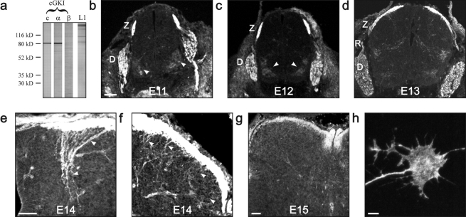
cGKIα is selectively expressed in developing DRG axons. (a) Western blot analysis demonstrates the presence of the α, but not the β, isoform of cGKI in embryonic DRGs. Lane 1, antibodies to both cGKI isoforms (c, common); lane 2, antibodies to the α isoform; lane 3, antibodies to the β isoform. For comparison, a blot of antibodies to L1 is shown. (b–h) Immunohistochemical detection of cGKI. (b–d) cGKIα distribution in the E11–E13 spinal cord. cGKIα is expressed in the DREZ and the developing dorsal funiculus as well as weakly and transiently on motoneuron columns (arrowheads) and in preganglionic neurons. Bar, 100 μm. (e) cGKIα expression in proprioceptive collaterals in the dorso-medial spinal cord at E14. Bar, 50 μm. (f) cGKIα expression in nociceptive collaterals in the dorso-lateral spinal cord at E14. (g) cGKIα distribution in the E15 spinal cord. Bar, 100 μm. (h) cGKI is distributed throughout a cultivated DRG growth cone. Bar, 5 μm. Sections were either stained by antibodies recognizing both isoforms of cGKI (E11, E12, and E13) or only the α isoform (E14 and E15). Both antibody preparations gave identical results. Tissue sections of cGKI-deficient mice did not stain with antibodies against cGKI (unpublished data). D, drg; R, dorsal root; Z, DREZ.

