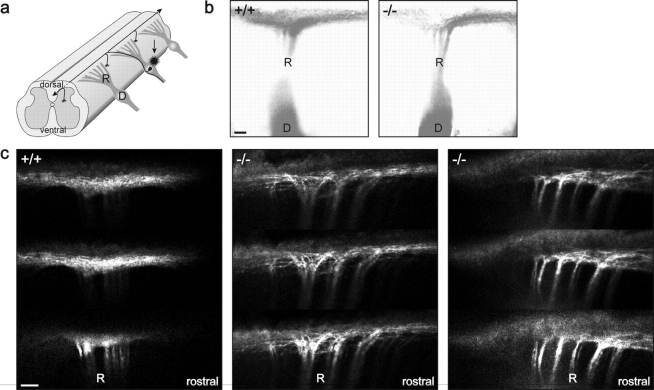
Figure 2.
Longitudinal growth of sensory axons is impaired in the spinal cord of cGKI-deficient mice. (a) Schematic drawing of the three-dimensional morphology of a single sensory neuron within the spinal cord. (b) To visualize the longitudinal branching, primary afferents in preparations of E12 or E13 embryos were labeled with the lipophilic axonal tracer DiI (Honig and Hume, 1989). Whole mounts were analyzed laterally with a fluorescence microscope. Whereas wild-type (+/+) DRG axons branch equally in rostral and caudal direction, cGKI-deficient (−/−) DRG axons have a strong preference for growing primarily in rostral direction (to the right). Fluorescence images were inverted. Bar, 100 μm. (c) Three adjacent confocal images of DiI-labeled axons of wild-type (+/+) and mutant mice (−/−) within the DREZ are shown. Note that mutant axons turn preferentially rostrally as bundles. Bar, 100 μm. D, DRG; R, dorsal root.

