
Figure 5.
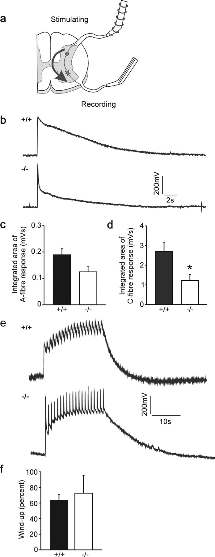
Impairment of the VRP of the spinal cord in the absence of cGKI. (a) In vitro–hemisected spinal cord preparation to measure monosynaptic and polysynaptic reflexes evoked by primary afferents. A scheme of a cross section of the spinal cord is shown and stimulating and recording electrodes are indicated. Dorsal is up. (b) VRPs of wild-type and cGKI-deficient neonatal mice. Electrical stimulation of dorsal root fibers generates a potential that can be recorded at the ventral root. The VRP is composed of a fast component evoked by A-fibers and a slower one activated by C-fibers (Heppenstall and Lewin, 2001). (c and d) Quantification of the VRP in wild-type and cGKI-deficient mice. The integrated area of the C-fiber area is significantly reduced in cGKI-deficient mice (P < 0.05; n = 6 per group). The integrated area of the A-fiber response was reduced but did not reach significance (P = 0.06). (e) Wind-up in wild-type and cGKI-deficient neonatal mice. Repetitive electrical stimulation of dorsal root fibers (20 times, 1 Hz) evokes an increase in the potential recorded from the ventral root. (f) Quantification of wind-up in wild-type and cGKI-deficient mice. Wind-up, measured as the percentage increase in the potential, is not different between wild-type and cGKI-deficient mice (P = 0.7; n = 6 per group).