Figure 6.

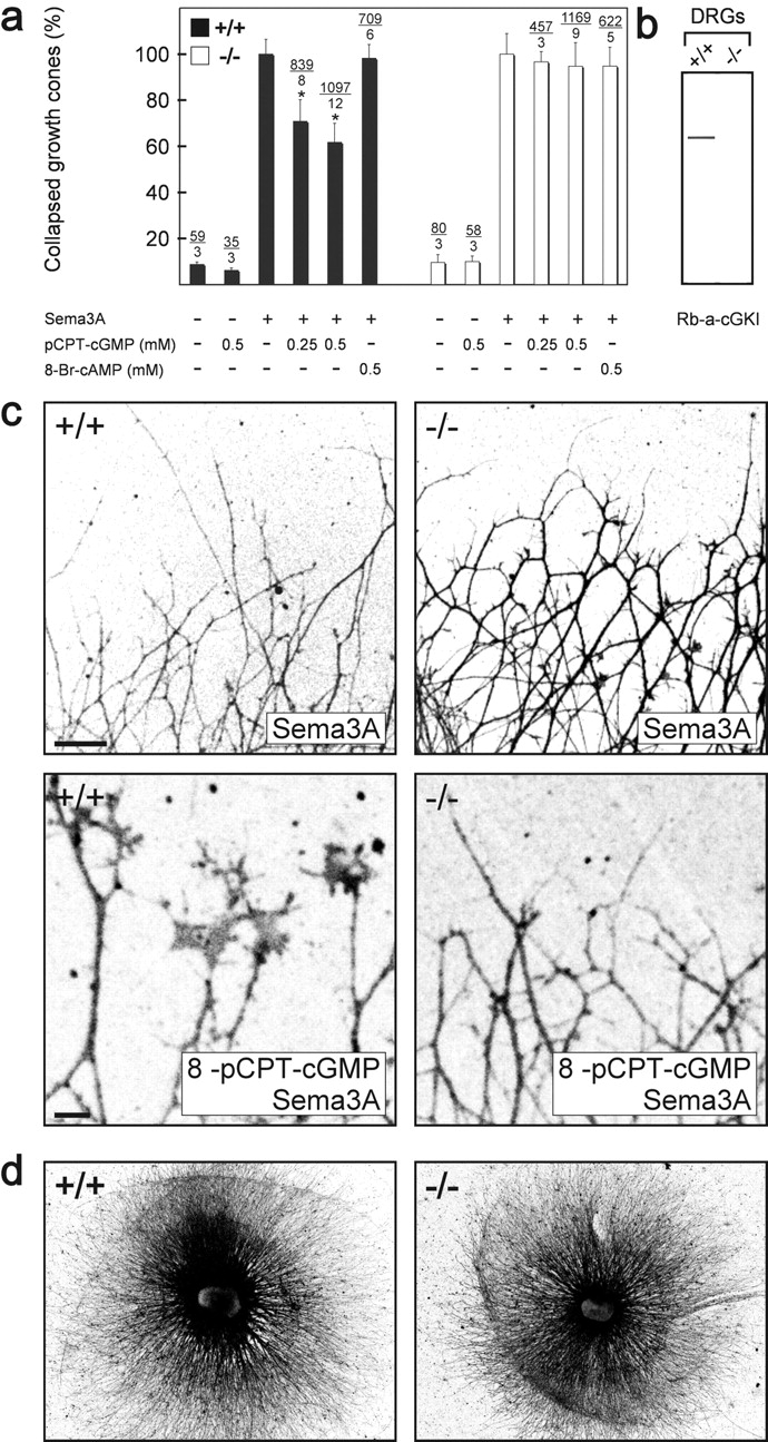
cGKI counteracts semaphorin 3A–induced growth cone collapse in vitro. (a) Growth cone collapse assay in the presence of 8-pCPT-cGMP or 8-Br-cAMP using DRG explants from litter-matched wild-type (+/+) or cGKI-deficient mice (−/−). The concentration and the presence of semaphorin 3A is indicated below the columns. The percentage of collapsed versus uncollapsed growth cones is presented and the number of counted collapsed growth cones and DRGs is given above the columns. The effect of semaphorin 3A alone was calculated as 100%. Error bars refer to SEM. Asterisk indicates significant difference from the samples without pretreatment with 8-pCPT-cGMP, P < 0.001 (t test). (b) cGKI protein could be detected in Western blots of DRG of wild-type (+/+) but not in DRGs of cGKI-deficient (−/−) embryos using polyclonal antibodies directed to both isoforms of cGKI. (c) Growth cones from wild-type (+/+) or cGKI-deficient (−/−) mice in the presence of semaphorin 3A alone or semaphorin 3A and 0.5 mM 8-pCPT-cGMP. Neurons were stained by mAb A2B5 for visualization and fluorescence images were inverted. DRGs from heterozygous (+/−) mice behaved as wild-type DRGs (unpublished data). Bar, 100 μm. Note that growth cones in the bottom images are shown at a higher magnification than in the top images. Bar, 10 μm. (d) Outgrowth of DRG explants of cGKI-deficient mice (−/−) is indistinguishable from those of wild-type mice (+/+). Explants from lumbar regions of litter-matched E13 embryos grown for 24 h on laminin-1 were silver stained (Rager et al., 1979).
