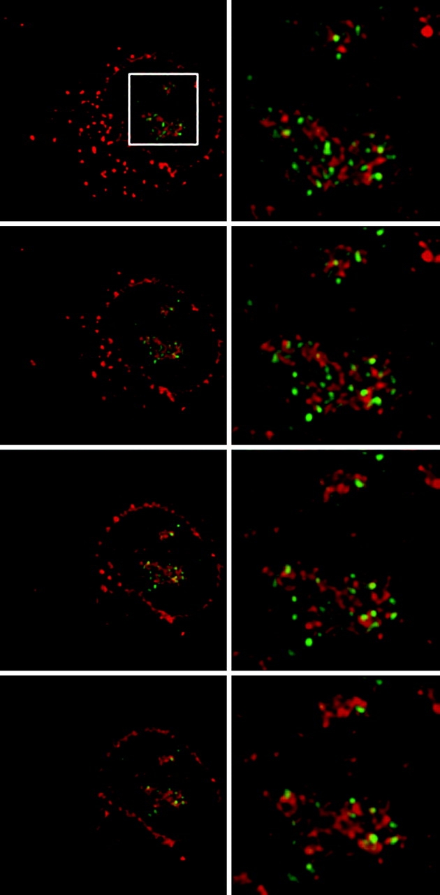
Figure 3.

Three-dimensional deconvolution shows minimal overlap between UBF and SRP RNA. Image stacks were captured and subjected to constrained interative deconvolution as described in Materials and methods. (Left) Successive vertical midplanes of SRP RNA signal (red) in a NRK cell immunostained for UBF (green). (Right) Corresponding midplanes enlarged from the boxed region (top left). Yellow color indicates areas of overlap between UBF and SRP RNA.
