Figure 2.
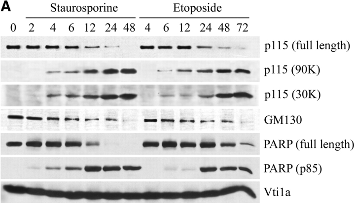
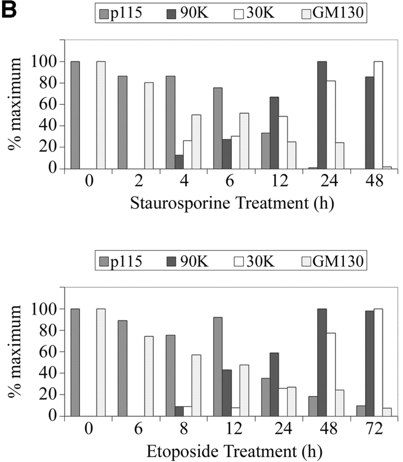
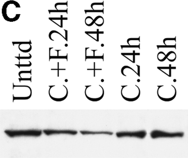
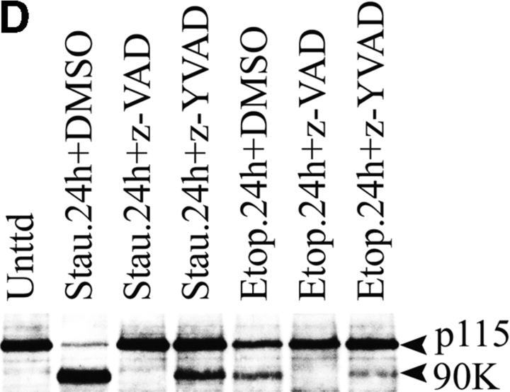
The levels of GM130 and p115 decrease during apoptosis. p115 is cleaved by caspases into two fragments. (A) COS7 cells were incubated with staurosporine or etoposide. At the indicated times, cell lysates were prepared for Western blotting using antibodies to GM130, p115, PARP, or the Golgi SNARE, Vtila. p115 antibodies revealed two polypeptides of 90 and 30 kD in apoptotic lysates. (B) Quantitation of GM130 and p115 breakdown and the appearance of the 90- and 30-kD polypeptides in staurosporine- and etoposide-treated cells. Note that the appearance of the 90- and 30-kD polypeptides coincided with the decrease in full-length p115, suggesting a precursor product relationship. (C) HeLa cells were treated with activating anti-Fas antibodies and cycloheximide (C.+F.). Cell lysates were prepared 24 or 48 h after treatment and analyzed by Western blotting using an antibody to p115. Unlike control samples treated with cycloheximide alone, the p115 levels of cells incubated with both anti-Fas antibodies and cycloheximide were decreased. (D) Lysates were prepared from COS7 cells treated with staurosporine or etoposide for the indicated times in the presence of z-VAD-fmk or z-YVAD-fmk. Lysates were analyzed by Western blotting using an antibody to p115. Generation of the 90-kD polypeptide in apoptotic cell lysates was inhibited in the presence of the apoptotic caspase inhibitor z-VAD-fmk.


