Figure 6.
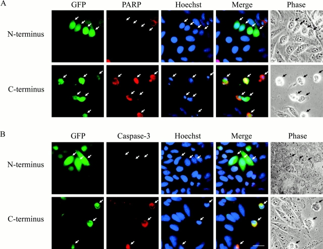
Expression of the COOH-terminal caspase cleavage fragment of p115 induces caspase activation and apoptosis. (A) HeLa cells were cotransfected with vectors expressing GFP and either the NH2- or COOH-terminal caspase cleavage fragment of p115. At 24 h after transfection, cells were processed for immunofluorescence microscopy and stained with Hoechst 33238 (blue) and a polyclonal antibody that recognizes only the p85 caspase cleavage fragment of PARP (red). Transfected cells were identified by GFP fluorescence (green). Cells expressing the p115 NH2-terminal cleavage fragment did not stain for cleaved PARP, had normal nuclear staining, and were morphologically indistinguishable from untransfected cells (top). In sharp contrast, cells expressing the COOH-terminal cleavage fragment of p115 were positive for cleaved PARP and displayed additional apoptotic features including chromatin condensation and cell shrinkage (bottom). (B) HeLa cells were transfected and processed as above. Fixed cells were stained with Hoechst 33238 (blue) and a polyclonal antibody that specifically recognizes the p18 subunit of activated caspase-3 but not procaspase-3 (red). Transfected cells (green) expressing the COOH-terminal (bottom), but not the NH2-terminal (top), caspase cleavage fragment of p115 stained for the activated caspase-3 antibody. Arrows indicate transfected cells. Bar, 10 μm.

