
Figure 2.
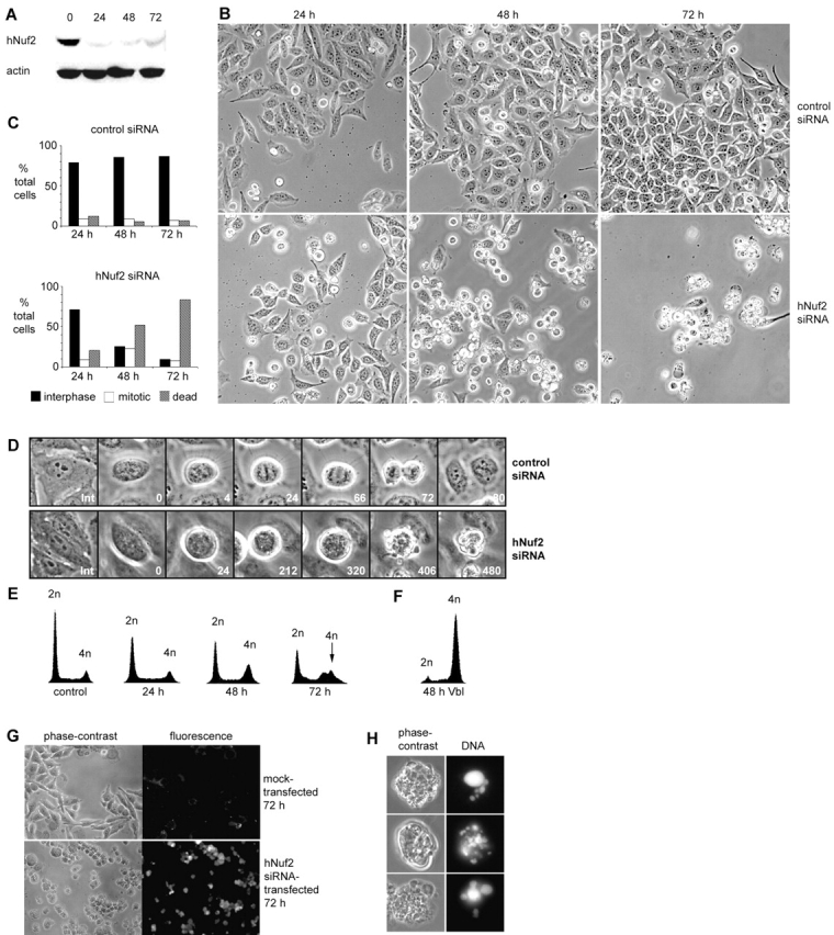
hNuf2-depleted cells exhibit a prolonged mitotic block and undergo cell death. (A) Reduction of hNuf2 levels after siRNA transfection. HeLa cells were transfected with hNuf2 siRNA, harvested 24, 48, and 72 h after transfection, and subjected to protein immunoblot analysis with hNuf2 antibodies and actin antibodies to control for gel loading. Time points are indicated above each lane, and time 0 indicates untransfected cells. (B) hNuf2 siRNA transfection results in a mitotic defect. Cells were transfected with either hNuf2 siRNA or a control 21-nucleotide siRNA duplex, and imaged 24, 48, and 72 h after transfection using a 10× phase objective. (C) Quantitation of control and hNuf2 siRNA–transfected cell phenotypes. Cells adherent to the culture dish with an intact nuclear envelope and decondensed chromatin were scored as interphase. Rounded cells with condensed chromosomes and smooth, uniform membranes were scored as mitotic. Cells that were multilobed, dense, and exhibited a nonuniform membrane were scored as dead (Mills et al., 1999; Zhang and Xu, 2002) (n = 6,000 cells per time point averaged from three independent experiments). (D) Mitotic progression of control siRNA– versus hNuf2 siRNA–transfected cells. 48 h after transfection, cells were transferred to live-cell chambers and time lapsed using a 40× phase objective. Top row, typical control siRNA–transfected cell; bottom row, typical hNuf2 siRNA–transfected cell. Time shown in minutes. (E) Time course of change in cellular DNA content after hNuf2 siRNA transfection. Time after transfection along the x axis is in hours, and untransfected cells were used as a control. Cells were analyzed by flow cytometry as described in the Materials and methods. (F) Cellular DNA content as analyzed by flow cytometry of cells treated for 48 h with 10 μM vinblastine. (G) Uptake of Trypan blue in hNuf2 siRNA–transfected cells. Mock-transfected and hNuf2 siRNA–transfected cells were incubated with Trypan blue 72 h after transfection and observed by phase-contrast and epifluorescence microscopy. (H) Nuclear fragmentation of hNuf2-depleted cells. Cells transfected with hNuf2 siRNA were fixed and stained with DAPI 48 h after transfection.