Figure 6.

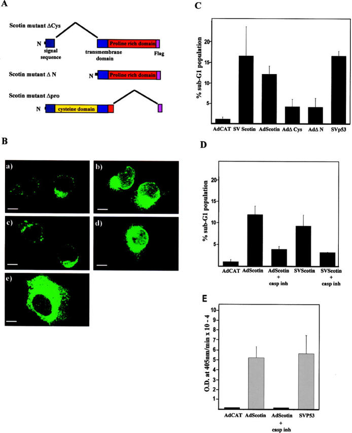
wt Scotin protein has an intrinsic proapoptotic activity. (A) Schematic representation of the Scotin mutants. (B) Scotin mutants deleted of the NH2 terminus are located in the ER. H1299 cells were transfected with 0.5 μg of AdScotin-FLAG (a), or SVScotin-FLAG (b), or AdΔCys (c, or AdΔN (d), or SVΔpro (e). Cells were stained with anti-FLAG (M2) mouse mAb followed by anti–mouse antibody conjugated to FITC. Bars, 5 μm. (C) wt Scotin protein induces apoptosis after transfection. The DNA content of each transfected population was determined by flow cytometry analysis. The percentage of sub-G1 DNA content represents the percentage of apoptotic cells 48 h after transfection. H1299 cells transfected with the following different expression vectors: 5 μg/ml AdCAT, or 10 μg/ml SVScotin, or 5 μg/ml AdScotin, or 2 μg/ml SVp53, or 5 μg/ml AdΔCys, or 5 μg/ml AdΔN. Histogram represents the average of at least three independent transfections. SDs are reported as error bars. (D) Scotin-mediated apoptosis is inhibited by caspase inhibitor. The DNA content of each transfected population was determined by flow cytometry analysis. H1299 cells transfected with 5 μg/ml of AdCAT, 5 μg/ml of SVScotin-FLAG, or 5 μg/ml of AdScotin-FLAG were incubated in presence or absence of the caspase inhibitor Z-VAD-FMK. Histogram represents the average of at least three independent transfections. SDs are reported as error bars. (E) Scotin induces caspase-3 activation. The caspase-3 activity was determined using the Colorimetric Caspase-3 Cellular Activity Assay kit. The H1299 cells were transfected with 5 μg/ml of AdCAT, 5 μg/ml of AdScotin-FLAG, or 2 μg/ml of SVp53 expression vectors. As indicated, cells were incubated in the presence or absence of the caspase inhibitor Z-VAD-FMK. Histogram represents the average of three independent transfections. SDs are reported as error bars.
