Figure 7.

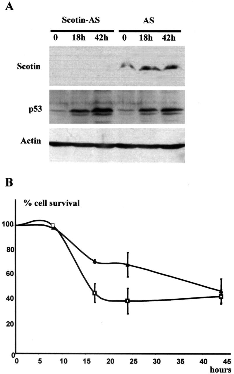
Scotin is involved in p53-dependent apoptosis. (A) Endogenous Scotin expression is inhibited in cells stably transfected with Scotin AS vector. 3T3 cells stably transfected with control antisense (AS) expression vector and 3T3 cells stably transfected with Scotin antisense (Scotin-AS) expression vectors were treated with actinomycin D (60 ng/ml). Proteins were extracted at the indicated time and analyzed by Western blot. Scotin expression was detected by using anti–mouse Scotin antibody. As a positive control, p53 induction was determined using CM5 anti–mouse p53 antibody. Protein loading was controlled by antiactin antibody. (B) Cells expressing a low level of Scotin protein are resistant to DNA damage. For the trypan blue exclusion assay, Scotin-AS cells (▴) or AS cells (□) were irradiated by UV-C (15 J/m2) and harvested at the indicated times. Viable cells were determined by trypan blue exclusion and counted using a hemocytometer. The percentage of cell survival was calculated as the number of viable cells in relation to the number of cells plated at the start of the trial. Each point is the average of at least three experiments. SDs are reported as error bars.
