
Figure 2.
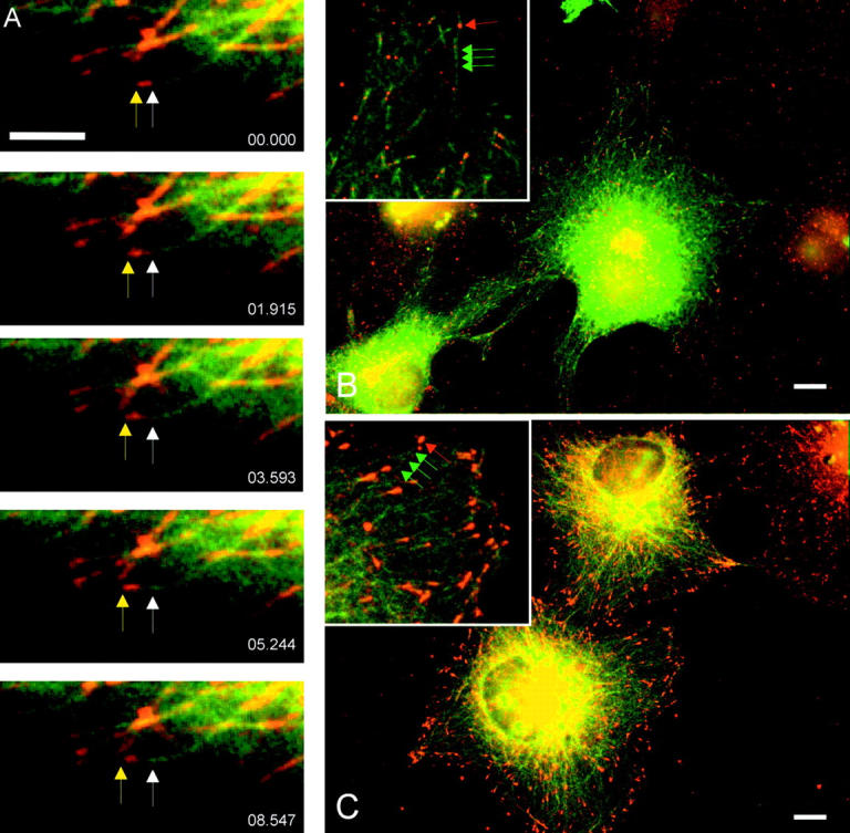
Microtubule plus-end–specific binding of p150 Glued. (A) Cells transfected with RFP-p150Glued and GFP-tubulin were subjected to live-cell imaging and analyzed as time-lapse movies. The white arrow shows the initial position of RFP-p150Glued while the yellow arrow shows the growing microtubule tip. Elapsed time of sequence is reported in lower right corner (s.ms). (B and C) Cells transfected with GFP-p150Glued 1–330 (green) were fixed with methanol and labeled by IFM for CLIP-170 (B, red) or EB1 (C, red). Insets show higher magnification of GFP-p150Glued (green arrows), CLIP-170 (red arrows), and EB1 (red arrows). Bars: (A) 5 μm; (B and C) 10 μm.