Figure 4.
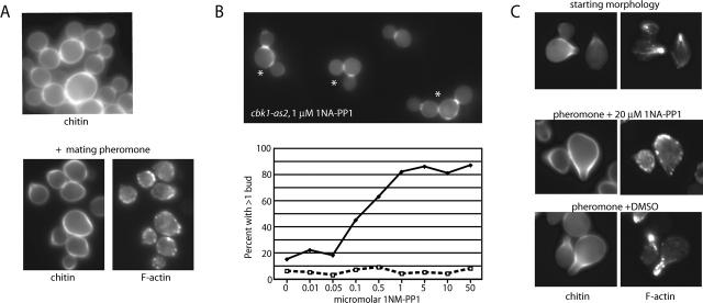
Inhibition of Cbk1p kinase activity affects cell separation and polarized growth. (A) Chitin and F-actin organization in cells carrying cbk1 D475A (FLY1007). (Top) Chitin staining of asynchronous cells, showing failure of mother/daughter separation and rounded cell shape. (Bottom) Chitin and F-actin organization in cbk1 D475A cells treated with pheromone for 120 min, demonstrating failure of mating projection formation. (B) Cell separation defects caused by 1NA-PP1 treatment of cbk1-as2 cells (FLY1008). The top panel shows dispersed cbk1-as2 cells treated with 1 μM 1NA-PP1 for 2 h; cells counted as instances of mother/daughter separation failure indicated by an asterisk. Graph indicates percentage of cells failing to separate after a 2-h treatment with varying concentration of 1NA-PP1. □, wild-type cells; ♦, cbk1-as2 cells. (C) Maintenance of mating projection growth after inhibition of Cbk1p. The top panels show mating projection formation by cbk1-as2 cells treated with mating pheromone (α-factor; note correspondence of F-actin polarization and chitin deposition) ∼80% of cells exhibited similar morphology (n = 100). The middle panels show a 40-min treatment of these cells with 20 μM 1NA-PP1 in continued presence of pheromone; ∼55% of cells exhibited a depolarized phenotype, ∼30% remained polarized, and ∼15% had not obviously formed mating projections. The bottom panels show control 40-min DMSO treatment of cells shown in top panels; no depolarization was evident.

