
Figure 5.
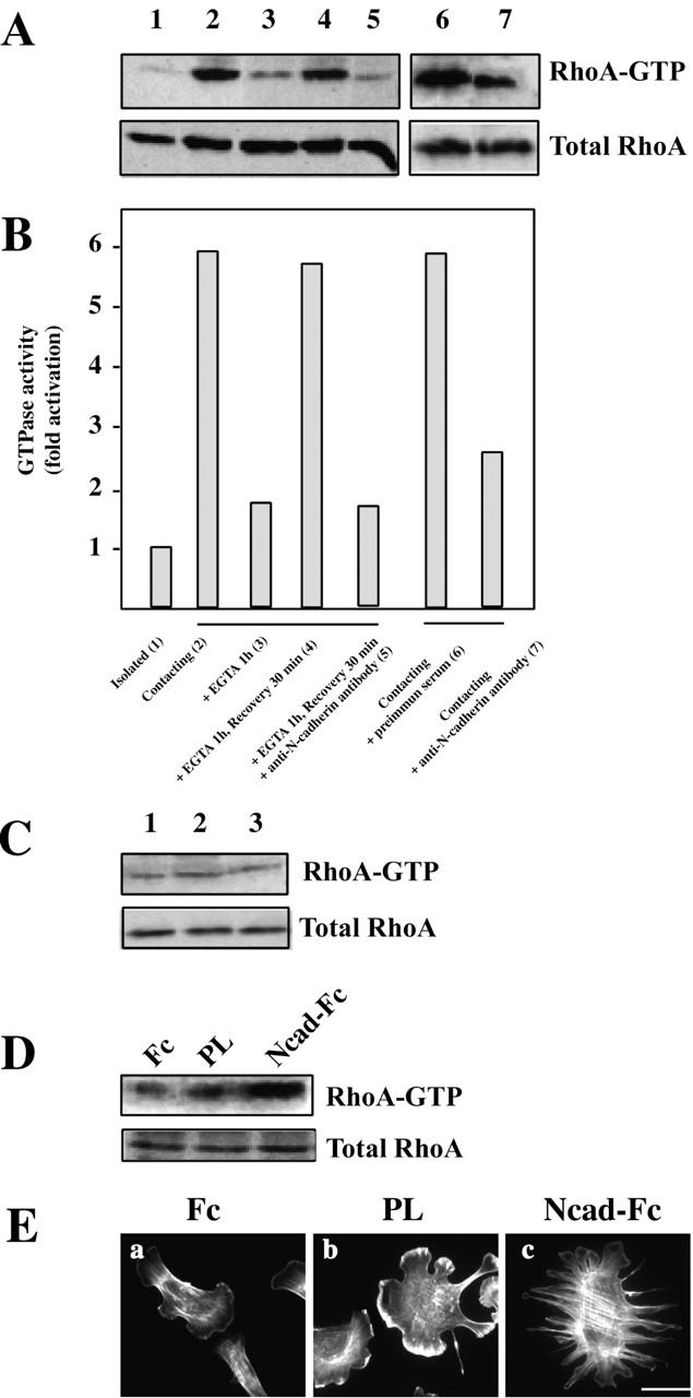
N-cadherin–dependent cell–cell contact formation activates RhoA GTPase. (A) The level of GTP-bound RhoA was measured using GST fused to the Rho-binding domain of the RhoA effector Rhotekin (GST-TRBD) in lysates obtained from cells treated as shown below the bars in B. RhoA molecules were detected by immunoblotting. (B) The results in A were analyzed by densitometry as described in Materials and methods. The histogram represents the GTPase activity normalized for the amount of total protein. Data are representative of more than three independent experiments. (C) The level of GTP-bound RhoA was measured in lysates obtained from isolated C2C12 myoblasts left untreated (1), treated with EGTA (2), and treated with EGTA before Ca2+ restoration (3). Data are representative of three independent experiments. (D) The level of GTP-bound RhoA was measured in lysates obtained 2 h after plating of C2C12 on surface coated with either Fc fragment, PL, or Ncad-Fc. Total and bound RhoA molecules are shown. Data are representative of three independent experiments. (E) C2C12 myoblasts plated onto surface coated with Fc fragment (a), PL (b), or Ncad-Fc (c) were stained for F-actin distribution with rhodamine-labeled phalloidin. Bar, 10 μm.