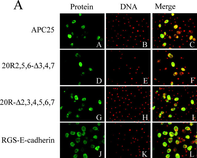
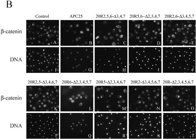
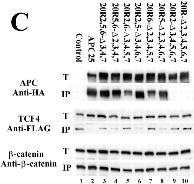
Figure 9.
Subcellular localization and complex formation with β-cat is altered in the presence of APC NES mutants. (A) Yeast cells with integrated β-cat and TCF4 in combination with integrated wild-type APC-25 (APC-25) or APC-25 NES mutants or expressing RGS–E-cad from a plasmid were subjected to IF with an antibody against the HA epitope (APC-25 proteins) or against E-cad. Proteins (Protein, panels A, D, G, and J) and DNA (DNA, panels B, E, H, and K) were photographed. False color images were captured and merged (Merge, panels C, F, I, and L), overlapping signals of protein (green) and DNA (red) are indicated by yellow color. (B) Yeast cells with integrated β-cat and TCF4 alone plus either a control vector (Control), wild-type APC-25 expression vector (APC-25), or expression vectors for the indicated APC-25 NES mutants were subjected to IF with an antibody against β-cat. Proteins (panels A–E and K–O) and DNA (panels F–J and P–T) were photographed. (C) β-cat immunoprecipitates of protein extracts from cells expressing β-cat and TCF4 (Control) and either wild-type APC-25 or APC-25 NES mutants were analyzed by Western blot. Antisera against the HA tag (to detect APC-25 proteins), FLAG tag (to detect TCF4), or β-cat were used to identify the corresponding proteins in total cell extracts (T) as well as in complex with β-cat (IP).


