Figure 6.
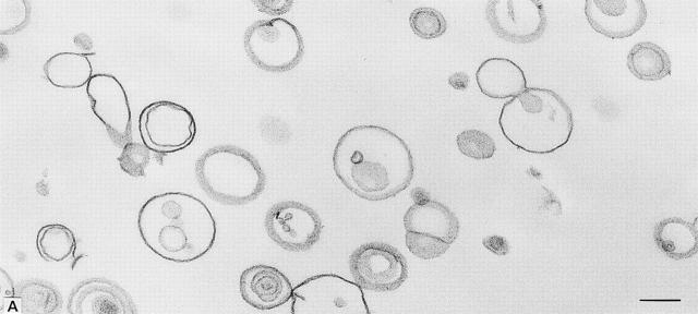
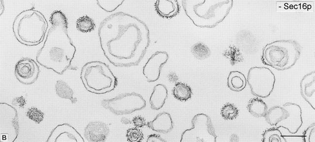
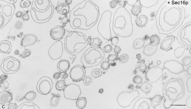
Thin-section electron microscopy of major–minor mix liposomes incubated with and without COPII proteins, GMP-PNP, and MBP–Sec16p. (A) No protein addition showing large, uncoated, uni- and multilamellar liposomes. (B) COPII and GMP-PNP promote coating, budding, and coated vesicle formation. (C) COPII, GMP-PNP, and Sec16p produce groups of vesicular profiles in close apposition to larger liposomes. Filamentous material not seen in B tethers liposomes and coated vesicles together. Bars, 0.2 μm.


