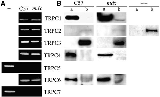
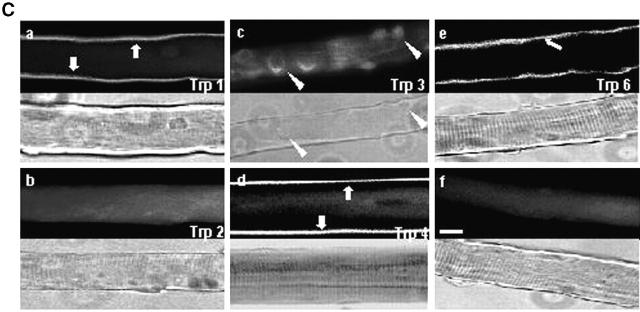
Figure 4.
Expression and localization of TRPC isoforms. (A) Detection of the presence of TRPC mRNA by RT-PCR in C57 and mdx fibers. Brain (+) has been used as a positive control to prove the absence of TRPC5 and TRPC7. (B) Localization of endogenously expressed TRPC1, 2, 3, 4, and 6 in C57 and mdx fibers. Triton-soluble (a) or Triton-insoluble (b) proteins were separated on 8% SDS-PAGE, transferred onto PVDF membrane, and processed for Western blot analysis with specific pAbs. Sol 8 cells (++) have been used as a positive control for TRPC2. (C) Localization of Trp1, 2, 3, 4, and 6 proteins in mdx muscle fibers immunostained with specific antibodies (f, staining with the secondary antibody as a negative control). The corresponding light micrographs are presented for each case. Bar, 10 μm.


