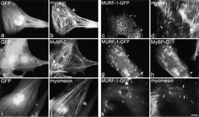
Figure 4.
Expression of GFP–MURF-1 also perturbs the organization of thick filament components. Transfected myocytes were stained with antibodies to myosin (b and d), MyBP-C (f and h), and myomesin (j and l). In many GFP–MURF-1–expressing myocytes, staining for thick filament components was perturbed (d, h, and l), compared with myocytes transfected with GFP alone (b, f, and j). Single arrows mark perturbed thick filament component staining, and double arrows mark regular, striated thick filament staining. Note, size and intensity of the GFP–MURF-1 cytoplasmic aggregates vary from cell to cell (c, g, and k, arrowheads). Bar, 10 μm.

