
Figure 7.
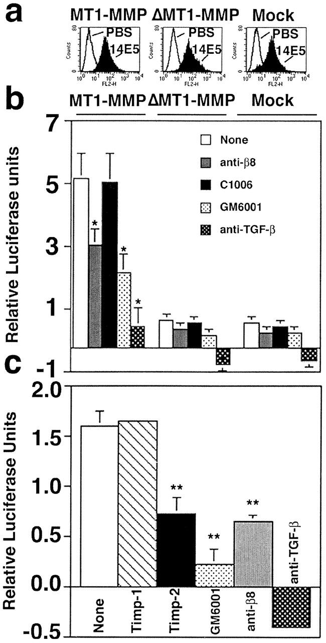
αvβ8 mediates activation of TGF-β in β8-overexpressing H1264 cells reconstituted with MT1-MMP activity. (a) Flow cytometry of β8-transduced MT1-MMP, ΔMT1-MMP, or mock-transduced H1264 cells demonstrates equivalent levels of surface expression of β8 using an anti-β8 antibody (14E5). Histograms using arbitrary units are shown. (b) β8-overexpressing H1264 cells transduced with either MT1-MMP, ΔMT1-MMP, or the retroviral vector alone (mock) (1.6 × 10⁴) were cocultured with TMLC (1.6 × 10⁴) reporter cells in the presence or absence of inhibitors: anti-β8 (37E1), control peptide (C1006), GM6001, or the pan–TGF-β1 antibody (1D11). Asterisks indicate significantly different than untreated MT1-MMP–expressing cells. (c) The endogenous inhibitor TIMP-2 but not TIMP-1 inhibits αvβ8-mediated activation of TGF-β in H1264s cells. β8-overexpressing, MT1-MMP–expressing H1264s cells were cocultured with TMLC in the presence or absence of TIMP-1 (1 μg/ml), TIMP-2 (1 μg/ml), GM6001 (5 μM), anti-β8 (37E1), or pan-TGF-β1 (1D11). Relative luciferase units are shown (light units of cocultured cells in the presence or absence of inhibitors minus light units of TMLC cells alone) in b and c. Negative luciferase values were occasionally observed due to a small background of TGF-β activation by the TMLC cells. Single and double asterisks indicate treated cells compared with untreated cells (*p < 0.01; **p < 0.001).