Figure 8.
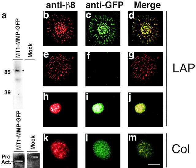
αvβ8 and MT1-MMP colocalize in substrate contacts. (a, top) Immunoprecipitation of MT1-MMP–GFP from 125I cell surface-labeled MT1-MMP–GFP–expressing HT1080 cells. The catalytically active 90-kD MT1-MMP–GFP fusion protein (asterisk) was immunoprecipitated with anti-GFP antibodies from MT1-MMP–GFP–expressing HT1080 β8 cells but not mock-transduced HT1080 β8 cells. The 70-kD MT1-MMP–GFP band represents a catalytically inactive degradation product. (a, bottom) Gelatin zymography of supernatants from MT1-MMP–GFP–transduced or mock-transduced HT1080 β8 cells. The migration of Pro (Pro−) and active (Act.−) forms of MMP-2 are shown. (b–m) Confocal images of immunofluorescence microscopy. β8-expressing, MT1-MMP–GFP–expressing HT1080 cells (b–d and k–m); β8-expressing HT1080 cells (e–g); GFP-expressing HT1080 cells (h–j). Cells were allowed to attach 4 h to LAP-β1 (10 μg/ml coating concentration)-coated slides. After fixation and permeabilization, colocalization of β8 and GFP was determined using polyclonal anti-β8 and monoclonal anti-GFP antibodies. Pseudocolored confocal images of β8 (red) and GFP (green) staining taken in the plane of the substrate are shown. Bar, 7.5 μM.

