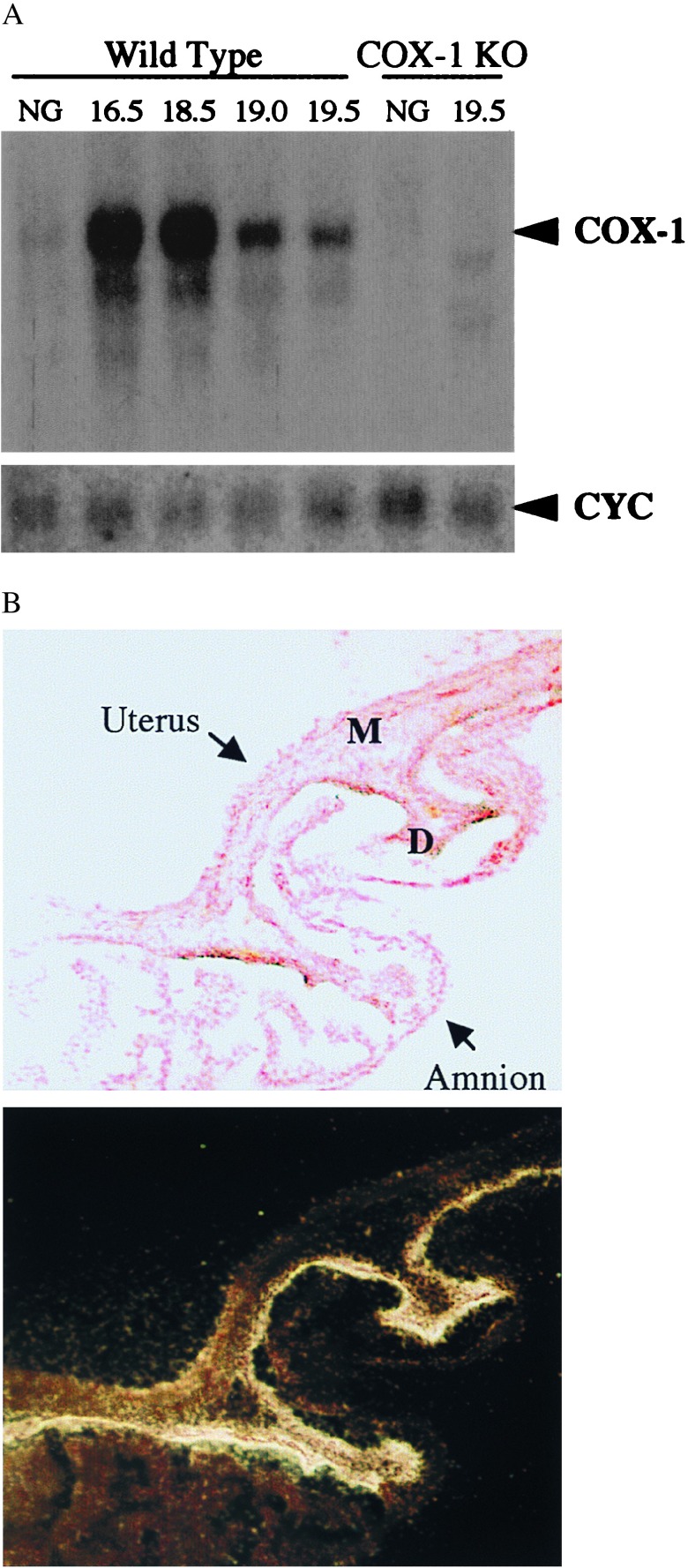
Figure 1.
Induction of COX-1 expression during gestation. (A) Northern blot analyses of total uterine RNA during pregnancy. Samples from nongravid (NG) or pregnant females of the gestational age in days given above the corresponding lanes were hybridized to COX-1 or cyclophilin A- (CYC) radiolabeled probes. (B) Decidual (endometrial) localization of COX-1 in gravid uterus at 18.5 d of gestation. Histologic sections of intact uterine implantation sites including uterus, fetus, placenta, and extra-embryonic membranes were hybridized to a radiolabeled COX-1 RNA probe. Shown is a representative bright field (Upper), nuclear fast red counterstained, region of uterus and amnion, along with the corresponding dark field photograph of the section (Lower), after exposure to emulsion and development. High level COX-1 expression is detected primarily within the decidua (D), as opposed to myometrium (M) or amnion, as demonstrated by deposition of silver grains.

