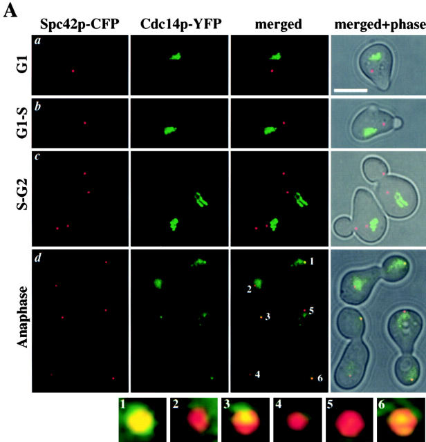
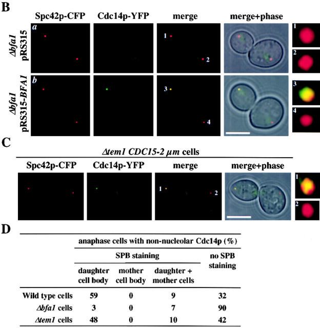
Figure 1.
Cdc14p binds to SPBs via Bfa1p. (A) Fluorescence microscopy of CDC14–YFP SPC42–CFP cells from an α-factor–synchronized culture: G1 (a), G1–S (b), S–G2 (c), and anaphase (d). Panels 1–6 on the bottom are magnifications of the SPBs in the anaphase cells shown above. (B) Cdc14p SPB localization is in part Bfa1p dependent. Fluorescence microscopy of anaphase cells from α-factor–synchronized CDC14–YFP SPC42–CFP Δbfa1 cells with plasmid pRS315 (a) or pRS315-BFA1 (b). Panels 1–4 are magnifications of the merged SPB signals. (C) Cdc14p SPB staining does not require Tem1p. Fluorescence microscopy of anaphase Δtem1 CDC15-2μ CDC14–YFP SPC42–CFP cells. Panels 1 and 2 are magnifications of the merged SPB signals. (D) Quantification of A–C. Cells in late anaphase with released Cdc14p (n > 100) were analyzed for Cdc14p–YFP SPB staining. Bars, 5 μm.


