
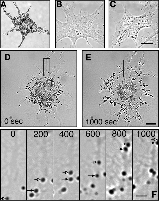
Figure 1.
Tracking organelles in cells with a limited number of melanosomes. (A) Cultured Xenopus melanophore. The large number of melanosomes makes tracking individual organelles impossible. (B) Cell after 4 wk of culture in 1 mM PTU. No melanosomes are seen in the cytoplasm. (C) Melanophore after 24 h recovery from PTU has a small number of melanosomes. (D and E) Melanophore after 24 h recovery from PTU before (D) and 1000 s after (E) addition of MSH, showing that the melanosomes in PTU-treated cells disperse in response to MSH. (F) Sequence of images taken during pigment dispersion of the cell shown in D and E. Arrows show positions of two melanosomes in subsequent frames. Time after addition of MSH (in seconds) is shown on each frame. Bright-field microscopy (A and D–F); phase–contrast microscopy (B and C). Bars: (A–E), 10 μm; (F) 2.5 μm.