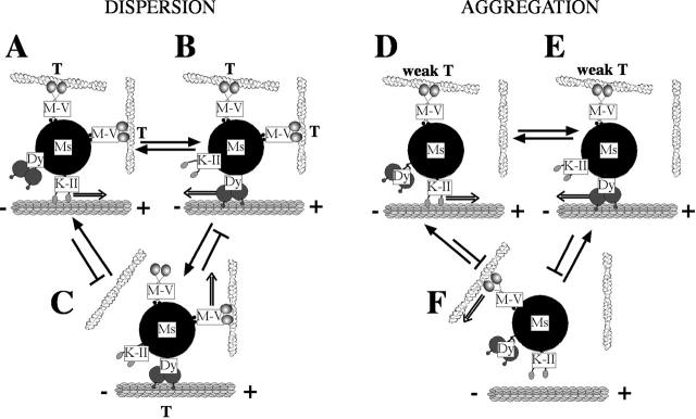
Figure 9.
Model for transport of melanosomes by actin- and microtubule-based motors. A pigment organelle can be moved in three different ways: along the microtubule by kinesin II (A and D) or dynein (B and E), or along the actin filaments by myosin V (C and F). Direction of movement is shown by double arrows. Although at any moment motion is dominated by one of the two transport systems (i.e., microtubules or actin), the motors of the other transport system are transiently active and interacting with their substrate (i.e., actin or microtubules). During dispersion, these transient interactions (indicated by T) are significant and allow myosin V to reduce the velocity of microtubule-based transport (A and B). The interactions also play a role in the switch between the two transport systems, that is, between B and C. Because myosin V activity predominantly decreases the length of minus end microtubule-based motion, we hypothesize that the switch from microtubule- to actin-based transport occurs only between state B and C (from dynein movement to myosin V movement) and that transitions from kinesin II movement (A) to myosin V movement (C) are rare. Because kinesin II but not dynein appears to win in tug-of-wars with myosin V, we suggest that the C to A transition is possible but not the C to B transition; however, we have no direct evidence on this point. During aggregation, myosin V activity is decreased, which results in reduced weak transient interactions (indicated by weak T) in D and E, and myosin V is no longer able to interfere with microtubule-based motion. Due to the weakness of the interactions, any time the microtubule motors are in contact with the microtubules they win the tug-of-war with myosin V and there is a transfer (F to D or F to E). Similarly, the reverse transfer from microtubules to actin-based transport (D to F or E to F) does not occur. M-V, myosin V; K-II, kinesin II; Dy, dynein; Ms,melanosome.

