
Figure 6.

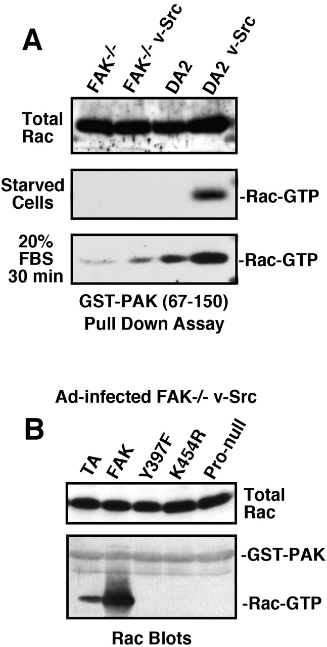
FAK activates Rac and serum stimulation promotes GFP-FAK localization to lamellipodia. (A) Rac activation was visualized in the indicated cells by pull-down assays using GST-PAK (67–150) followed by Rac blotting. Total Rac expression is shown in whole cell lysates. (B) FAK−/− v-Src cells were infected with Ad-TA or the indicated Ad-FAK constructs, and Rac activation was evaluated by GST-PAK binding. (C) FAK−/− v-Src cells were transfected with GFP-FAK, starved, and then stimulated with 10% FBS. Time-lapse confocal video-microscopy at 1-min intervals was obtained over 3 h (Video 1 available at http://www.jcb.org/cgi/content/full/jcb.200212114/DC1). Panels show a 9-min time series where GFP-FAK is stably localized to focal contacts and transiently recruited to lamellipodia extensions (arrows).