
Figure 4.
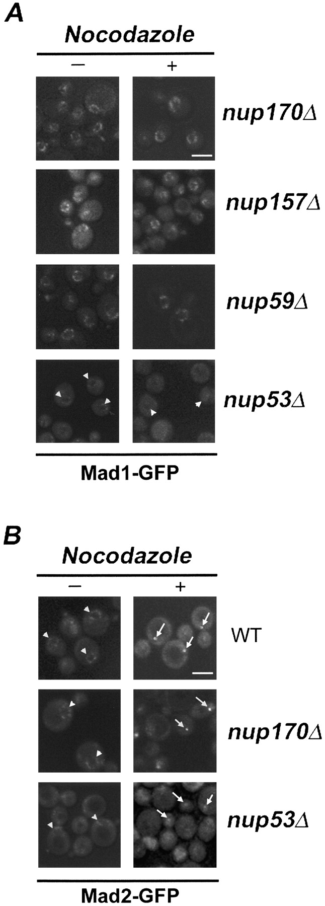
The effects of nup mutations on the subcellular distribution of the Mad1p–Mad2p complex. (A) The subcellular localization of Mad1-GFP in logarithmically growing (−) or nocodazole-treated (+) nup170Δ, nup157Δ, nup59Δ, and nup53Δ strains was examined using confocal fluorescence microscopy. (B) The localization of Mad2-GFP in logarithmically growing (−) or nocodazole-treated (+) WT, nup170Δ, and nup53Δ strains was examined using confocal fluorescence microscopy. Note, arrowheads point to the NE and arrows point to kinetochore signals. Bars, 5 μm.