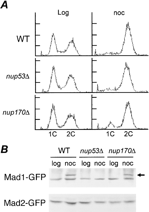
Figure 7.
Requirement of Nup53p for hyperphosphorylation of Mad1p. (A) nup170Δ and nup53Δ strains arrest in G2/M after treatment with nocodazole. FACS® analysis was performed on logarithmically growing (log) and nocodazole (noc)-treated (1.5 h) WT, nup53Δ, and nup170Δ strains. The positions of 1C and 2C DNA peaks are indicated. (B) Mad1p is not hyperphosphorylated in a nup53Δ strain. Logarithmically growing WT, nup53Δ, and nup170Δ strains expressing either Mad1-GFP or Mad2-GFP were treated with (noc) or without (log) nocodazole for 1.5 h. Total cell lysates were analyzed by immunoblotting using an anti-GFP antibody. The position of hyperphosphorylation Mad1-GFP is indicated by an arrow.

