
Figure 4.
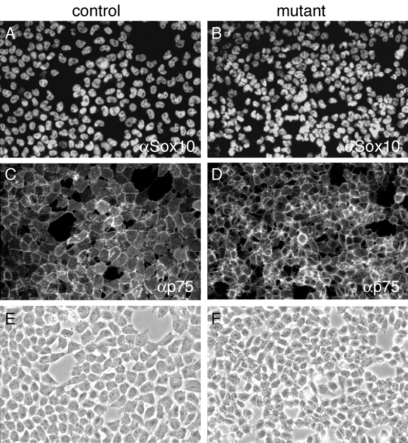

Normal emigration of mutant neural crest cells. Neural crest explants were obtained from neural tubes that had been isolated from control and β-catenin mutant mice at E9 and cultured for 20 h to allow emigration of neural crest cells. After emigration, neural crest cells were fixed and immunolabeled with anti-sox10 antibody (visualized by Cy3 fluorescence) (A and B) and anti-p75 antibody (visualized by FITC fluorescence) (C and D). Note that virtually all neural crest cells were double positive for the neural crest stem cell markers p75 and sox10. (E and F) Corresponding phase contrast pictures. (G) To compare and quantify the extent of control and mutant neural crest outgrowth after 20 h, the migration index was calculated using the NIH image 1.62 software (Materials and methods). Two independent experiments using nonsibling embryos were performed, scoring three explants of control and mutant embryos per experiment. Each bar represents the migration index (mean ± SD) of three different explants. Note that mutant explants were not significantly reduced in size and density.