
Figure 2.
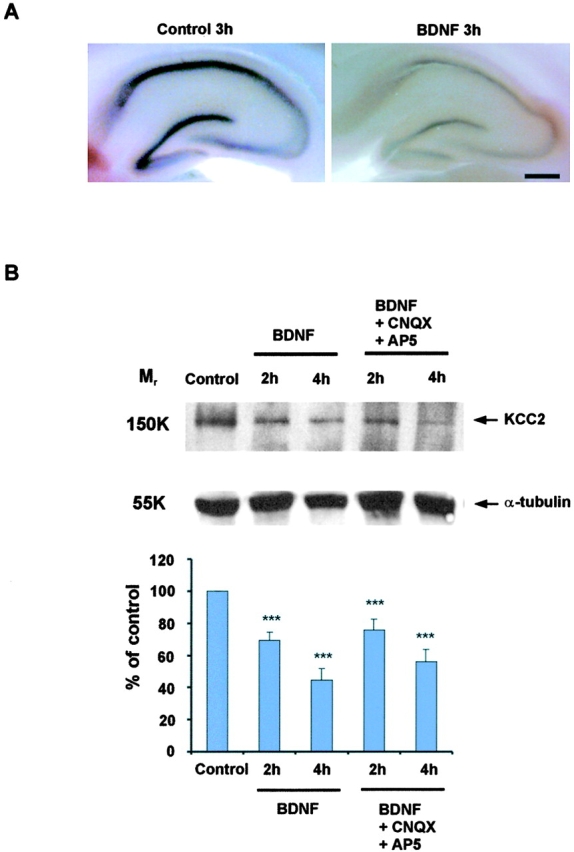
Rapid BDNF-induced down-regulation of KCC2 in acute hippocampal slices. (A) Free-floating in situ hybridization of acute hippocampal slices exposed to 100 ng/ml BDNF showing decreased KCC2 mRNA expression in all hippocampal regions as compared with control. (B) The top panel shows a representative Western blot of KCC2 expression at different time points after adding 100 ng/ml BDNF to the extracellular solution in the absence or presence of glutamate antagonists (CNQX and AP5). Normalized optical densities are shown in the bottom panel (n = 5; ***, P < 0.001 as compared with control using the t test). Note that the KCC2 protein is already down-regulated after 2 h. Bar, 1 mm.