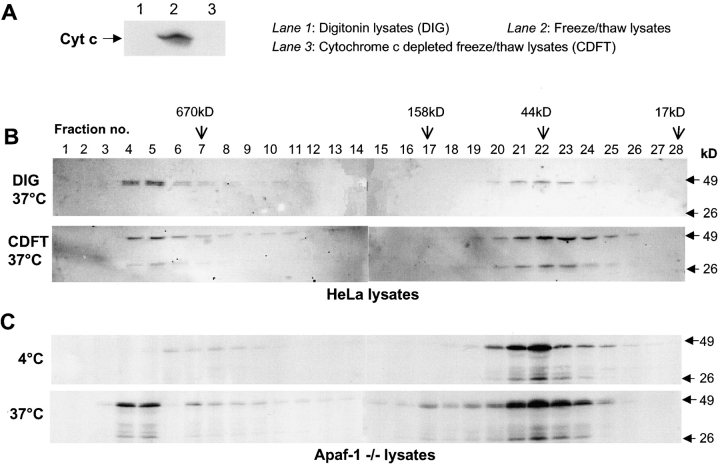
Figure 4.
Formation of caspase-2 complex is independent of cytochrome c and Apaf-1. In A and B, HeLa cell lysates were prepared by digitonin lysis or freeze/thawing. Immunoblotting in A, lane 1, demonstrated that digitonin lysates do not have any detectable cytochrome c (Cyt c). Cytochrome c from the freeze/thaw lysates was removed by immunodepletion. As shown in A, lane 3, immunodepleted cell extracts do not have any detectable cytochrome c. In B, digitonin lysates (DIG) and cytochrome c–depleted lysates (CDFT) were incubated at 37°C for 60 min, subjected to size exclusion chromatography, and analyzed by immunoblotting using a caspase-2 antibody. (C) Lysates from Apaf-1−/− cells were incubated at 4 or 37°C for 60 min and subjected to size exclusion chromatography. Fractions were precipitated with 10% TCA/0.07% β-mercaptoethanol/acetone at −20°C. Protein pellets were washed with cold 0.07% β-mercaptoethanol/acetone and resuspended in 1× SDS protein loading buffer. The entire sample was resolved by SDS-PAGE and analyzed by immunoblotting with the caspase-2 antibody.

