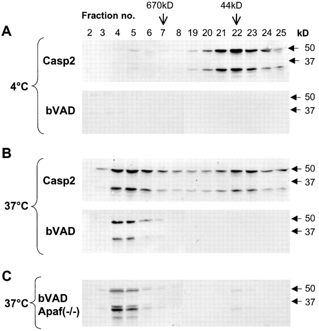
Figure 5.
Recruitment of caspase-2 to the large complex results in its activation. Lysates from HeLa (A and B) or Apaf-1−/− (C) cells were subjected to size exclusion chromatography. Aliquots from fractions were analyzed by immunoblotting using caspase-2 antibody (Casp2) or 200 μl of the HeLa fractions, or all of the Apaf-1−/− fractions were incubated with biotin–VAD-fmk (bVAD) for 60 min at RT followed by overnight incubation with streptavidin-Sepharose. Sepharose pellets were washed, and caspase-2 was detected by immunoblotting. HeLa lysates incubated at 4°C for 60 min (A); HeLa lysates incubated at 37°C for 60 min (B); Apaf-1−/− lysates incubated at 37°C for 60 min (C). Only caspase-2–containing fractions (2–8 and 19–25) are shown.

