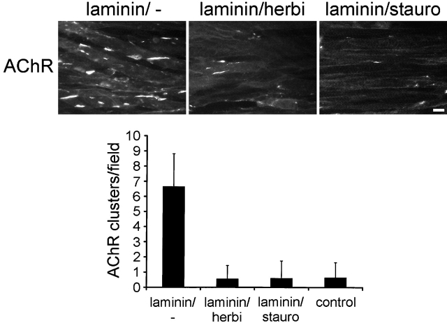
Figure 8.
Laminin-induced AChR clusters are dispersed by herbimycin and staurosporine. C2 myotubes were treated with 100 nM laminin-1 for 16 h and washed. Cells were reincubated without laminin and with or without inhibitors for 8 h as indicated. AChRs were visualized with rhodamine-α-btx and clusters quantitated. Data represent mean ± SD of 20 visual fields. Bar, 20 μm.

