
Figure 4.
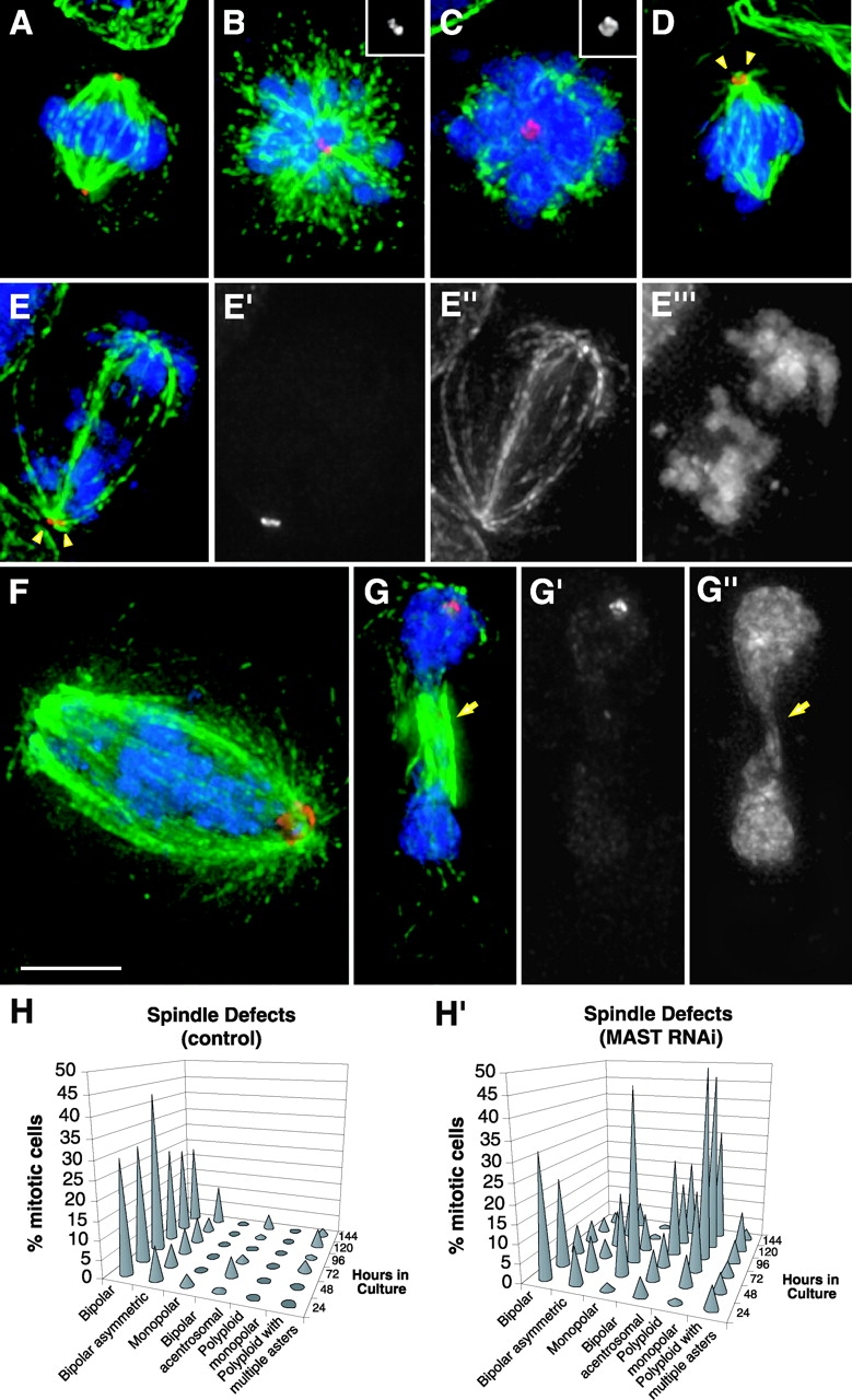
Organization of the mitotic apparatus after MAST RNAi. Cells after MAST RNAi were stained with an anti-CP190 antibody to reveal the centrosomes (red), an anti–α-tubulin antibody to visualize the microtubules (green), and DAPI to counterstain the DNA (blue). (A) Normal bipolar spindle in a control cell. (B and C) Monopolar and polyploid monopolar spindles with two and at least four centrosomes, respectively, observed 72 or 96 h after MAST RNAi. Centrosome staining alone can be seen in the insertion on top right of the figures. (D) Bipolar spindle with two centrosomes (arrowheads) only in one pole. (E) Anaphase-like cell displaying a bipolar spindle with centrosomes in a single pole and chromosomes distributed in two distinct populations on each side of the spindle. Unmerged images revealing the centrosomes (E′), spindle (E′′), and chromosomes (E′′′) are shown. (F) A cell with a bipolar spindle and multiple centrosomes clustered in a single pole where the chromosomes appear distributed along the spindle. (G) Abnormal telophase-like cell showing the formation of a cleavage furrow (arrows), with centrosomes in only one pole and decondensed chromatin. Separate centrosome and DNA staining can be seen in (G′) and (G′′), respectively. Bar, 5 μm. (H and H′) Quantification of the observed spindle defects in control and MAST-depleted cells, respectively (n > 100).