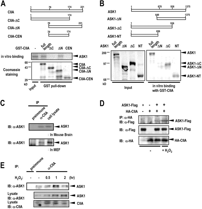
Figure 3.
CIIA physically interacts with ASK1. (A) 35S-Labeled ASK1 was produced by in vitro translation and incubated at 4°C for 3 h with GST-fused CIIA variants immobilized on glutathione-agarose beads. The bead-bound proteins were eluted and analyzed by SDS-PAGE and autoradiography. A lower part of the polyacrylamide gel was cut and stained with Coomassie brilliant blue to show the amount of GST-fused CIIA variants bound on the beads. (B) Binding of in vitro–translated 35S-labeled ASK1 variants to GST-CIIA was examined as in A. In A and B, the input 35S-labeled proteins (10%) were also shown. (C) The soluble fraction of mouse brain tissue or MEF homogenates was precleared with rabbit preimmune IgG and subjected to immunoprecipitation (IP) with rabbit anti-CIIA antibody, or rabbit preimmune IgG. The resulting precipitates were subjected to SDS-PAGE and analyzed by immunoblotting (IB) with anti-ASK1 antibody. Immunoblotting of cell lysates (5% of total) with anti-ASK1 antibody was also shown. (D) 293T cells were transfected with expression vectors encoding ASK1-Flag and HA-CIIA as indicated. After 48 h of transfection, the cells were untreated or treated with 500 μM H2O2 for 1 h. Cell lysates were subjected to immunoprecipitation with anti-HA antibody, and the resulting immunoprecipitates were subjected to immunoblot analysis with anti-Flag antibody. Cell lysates were also subjected to immunoblot analysis with the indicated antibodies. (E) L929 cells were untreated or treated with 500 μM H2O2 for indicated time periods. Cell lysates were subjected to immunoprecipitation and the resulting immunoprecipitates were analyzed by immunoblotting as in C. Cell lysates (5% of total) were also subjected to immunoblot analysis with anti-ASK1 or anti-CIIA antibody.

