
Figure 1.
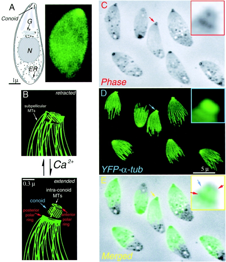
Drawing and LM images of Toxoplasma gondii. (A) (left) Drawing of T. gondii. showing the nucleus (N), Golgi (G), endoplasm reticulum (ER), and conoid. (right) Fluorescence image of living YFP-α-tubulin transgenic T. gondii. Intact cells appear uniformly green due to cytoplasmic YFP-α-tubulin. In extracted parasites (C–E), individual microtubules are visible. (B) Cartoon of conoid movement, modified from (Morrissette, 1995). In extracellular parasites, the conoid alternates between the retracted and extended states. Increase in cytoplasmic [Ca2+] induces extension. (C) Phase contrast image of A23187 treated YFP-α-tubulin transgenic T. gondii, some having extended conoids (red arrow). Two phase dark regions in the extended conoid (inset) correspond to the upper and lower polar rings (see B). (D) The brightly fluorescent apical end of YFP-α-tubulin transgenic parasites (blue arrow) protrudes together with the conoid (C, red arrow). (E) Merged phase contrast and fluorescence images. The apical spot of YFP fluorescence colocalizes with the phase light region (inset, blue arrow).