
Figure 4.
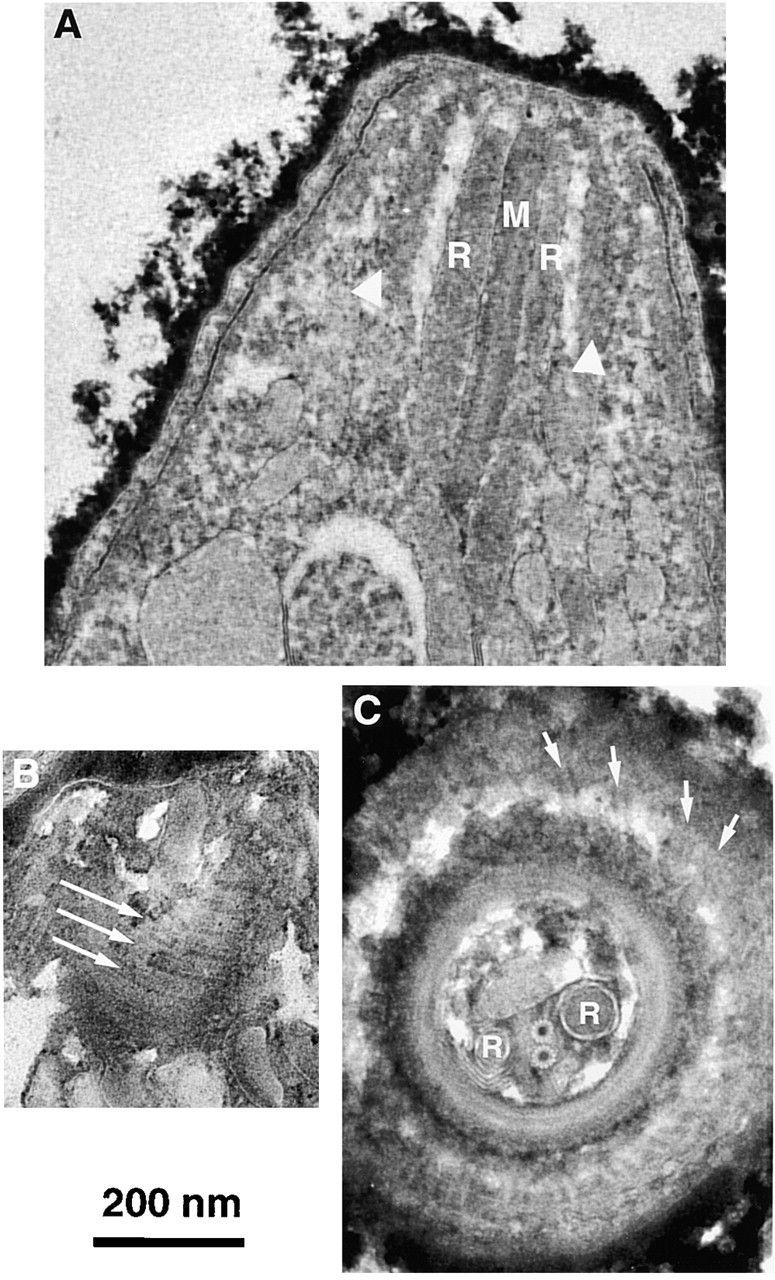
EM images of sections through the apical end of T. gondii with conoid retracted. (A) Sagittal section showing intraconoid microtubules (M), rhoptries (R) and walls of the conoid (white arrowheads). (B) A parasagittal section that grazed the wall of the conoid, revealing several of the spirally wound conoid fibers (long arrows). Faint longitudinal striations along each fiber can be seen. (C) Cross section of the apical end. The conoid appears as a circle of electron dense material with spiral striations. Just outside the conoid is a portion of the lower polar ring, to which the subpellicular microtubule (arrows) attach. Inside the conoid lie two rhoptries (R) positioned on either side of the two intraconoid microtubules.