
Figure 5.
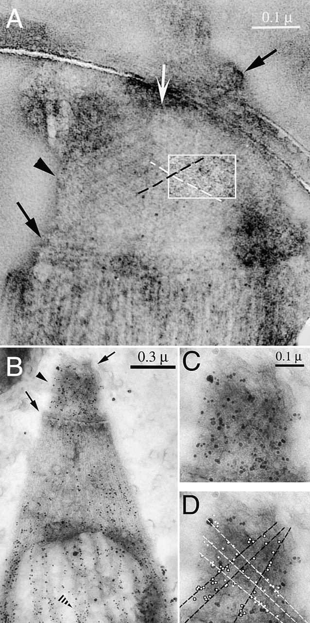
Immunogold labeling of T. gondii using anti-tubulin antibodies. (A) EM image of a negatively stained conoid of T. gondii, suspended over a hole in a carbon film. White arrow, intra-conoid microtubules; black arrows, upper and lower polar rings; black arrowhead, conoid region. Each conoid fiber is outlined by a dark line of electron-dense stain deposited in the groove between the fibers. The region within the white rectangle has been filtered and displayed with enhanced contrast to increase the visibility of the fibers and their longitudinal striations. (B) Immunogold labeling of T. gondii using a mixture of anti–α-tubulin and anti–β-tubulin antibodies. Black arrows and arrowheads as in A; striped arrowhead, a subpellicular microtubule, heavily decorated with gold particles. (C and D). Enlarged view of the conoid region from B. Most gold particles lie along conoid fibers, as indicated by the black and white lines in D.