
Figure 4.
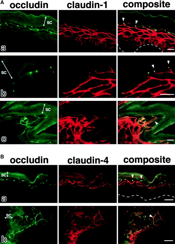
Occludin, claudin-1, and claudin-4 in the wild-type epidermis. (A) Double immunofluorescence microscopy of frozen sections of skin from the back of wild-type mice with antioccludin mAb and anti–claudin-1 pAb. In transverse sections (a and b), occludin was concentrated as dots in the most apical region of the lateral membranes of the granular cells in the second layer (arrowheads). This dot-like concentration of occludin was also found in some but not all of the granular cells in the first (uppermost) and/or third layer. Claudin-1 was distributed diffusely throughout plasma membranes of keratinocytes from stratum basale to granulosum, although the signal from the first layer of stratum granulosum was fairly weak (a). At higher magnification (b), in the lateral membranes of granular cells in the second layer claudin-1 was coconcentrated as dots together with occludin in the most apical regions (arrowheads). In thicker oblique sections (c), claudin-1 was coconcentrated precisely at the occludin-positive lines (arrowhead). SC, stratum corneum. The broken line represents the dermis/ epidermis border. Bars, 10 μm. (B) Double immunofluorescence microscopy of frozen sections of skin from the back of wild-type mice with antioccludin mAb and anti–claudin-4 pAb. Distinct from claudin-1, claudin-4 was distributed mainly in the second/third layers of stratum granulosum (a). Claudin-4 was also concentrated at the occludin-positive lines circumscribing granular cells (arrowhead) (b). SC, stratum corneum. The broken line represents the dermis/epidermis border. Bars, 20 μm.