
Figure 9.
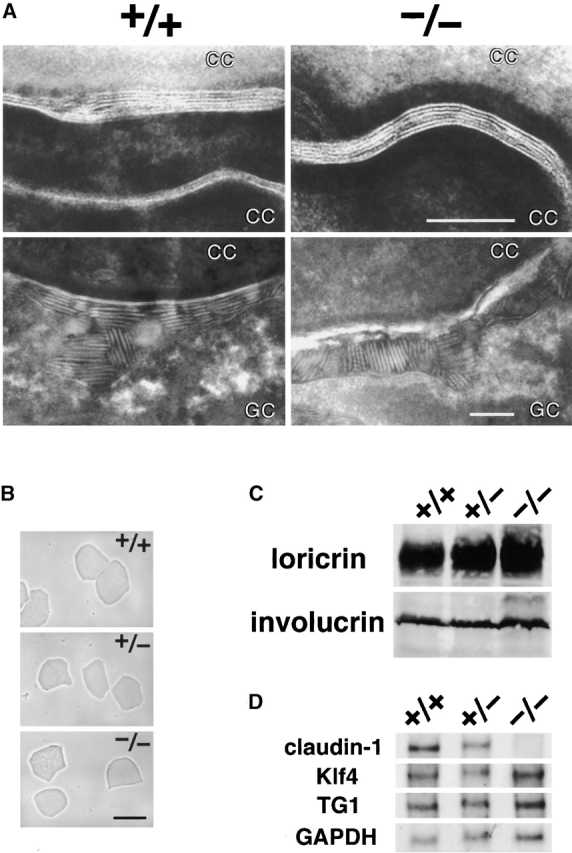
Stratum corneum in claudin-1–deficient mice. (A) Ultrathin section electron microscopic images of ruthenium tetroxide-stained stratum corneum of the skin. Both in the Cln1 + / + and Cln1 − / − epidermis, well-organized lipid lamellae and lamellar bodies were clearly observed between flattened cornified cells (CC; top) and between cornified cells and granular cells (GC; bottom), respectively. Bars, 0.1 μm. (B) Isolated cornified CEs. There was no clear difference in appearance between the Cln1 + / +, Cln1 + / −, and Cln1 − / − skin. Bar, 40 μm. (C) Expression levels of major components of cornified CEs, loricrin and involucrin, detected by immunoblotting. Whole cell lysates of the Cln1 + / +, Cln1 + / −, and Cln1 − / − skin were examined using specific antibodies. (D) Expression levels of Klf4, transglutaminase-1 (TG1), and GAPDH (control) detected by Northern blotting.